


How Tissues Are Processed
 Fixation — Tissue removed at biopsy is placed in a solution that quickly stops biochemical processes. Formalin has proven to be the most effective agent for stopping these processes and preserving the tissue’s overall appearance. “Fixation” is the term used to describe the desirable actions of formalin on tissues. Other agents (e.g., isopropyl alcohol) will fix tissues but induce artifacts that may seriously damage their diagnostic value.
Formalin is a 10% solution of aqueous formaldehyde. A wide-mouth unbreakable container of formalin is provided with each biopsy kit. Upon removal at biopsy, tissue is dropped directly into the provided solution ensuring immediate fixation.
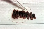
![]() Gross Examination — Upon receipt at our laboratory, fixed tissue is removed from the container and examined by one of our pathologists. The pathologist measures the tissue and describes its outer appearance then makes appropriate sections through the specimen describing cut-surfaces along the way. (These descriptions are included in the final biopsy report). Tissue sections are then aligned in a manner most advantageous for ultimate viewing; this alignment is often maintained by addition of gelatin.
Gross Examination — Upon receipt at our laboratory, fixed tissue is removed from the container and examined by one of our pathologists. The pathologist measures the tissue and describes its outer appearance then makes appropriate sections through the specimen describing cut-surfaces along the way. (These descriptions are included in the final biopsy report). Tissue sections are then aligned in a manner most advantageous for ultimate viewing; this alignment is often maintained by addition of gelatin.
Tissues containing hard substances need to be decalcified before gross sectioning and before preparation for microscopic diagnosis. Such calcified tissues are placed in decalcifying solution until the tissues can be cut with a scalpel (days to weeks). If sufficient, soft tissues may be detached from calcified structures and sent for immediate processing. Decalcified tissues are sent along when decalcification is complete.
![]()
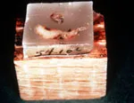 Paraffin Embedding — In order for tissues to be examined under the microscope, they must be cut into exceedingly thin sections. These sections need to be thinner than 7 micrometers (a micrometer = 1/1000th of a millimeter) to be viewed by transmitted light at magnifications up to 1000x. Infiltrating and imbedding the gelatin-aligned tissue sections in paraffin accomplish this. Tissue water is then removed by immersing the sections in alcohol solutions of increasing concentration. After dehydration, tissues are placed in xylol, a chemical compatible with alcohol and paraffin. After this preparation, the tissues are placed in a mold and then infiltrated with warm paraffin. Upon cooling, the tissues now reside in a paraffin block.
![]()
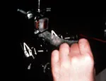 Thin Sectioning — After trimming, the paraffin block is mounted in a specialized cutting machine known as a “microtome.” A microtome is very heavy in order to dampen vibrations. It has a heavy and exceedingly sharp fixed blade. By turning a large wheel, the paraffin block is brought down across the cutting edge producing a thin section. After each wheel turn the block is advanced a few microns and another cut is made. During cutting the section is heated (by friction) just enough to attach the current one to the previous one producing a ribbon. Each of these sections contains very thin slices through the tissue to be studied. The paraffin block with the remaining tissue has an unlimited life span and is stored for future use.
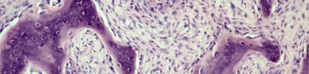
Transfer to Glass Slide — The paraffin ribbon is then floated on the surface water in a heated water bath. The water creates a flat surface and the heat allows for the sections to be separated from one another. A thin piece of glass (1″ x 4″ microscope slide) is placed in the water and a separated section is floated on it. This is repeated until sufficient sections are floated on slides. The tissue sections are examined; those with defects are discarded. When enough defect-free sections are affixed, the slides are placed in an incubator to dry, affix, and further flatten the tissue sections.
![]()
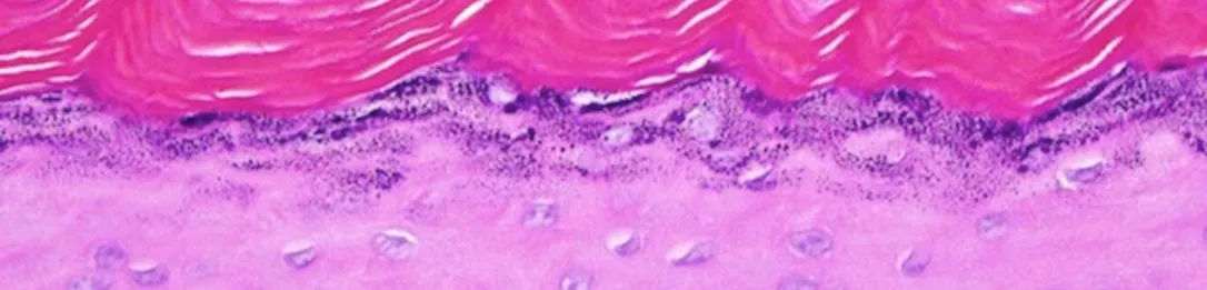
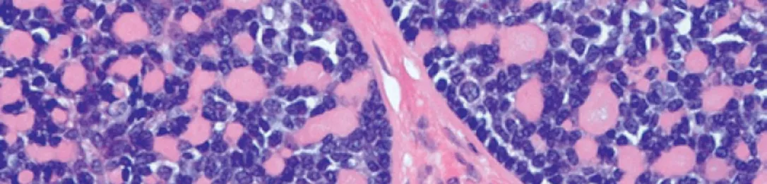
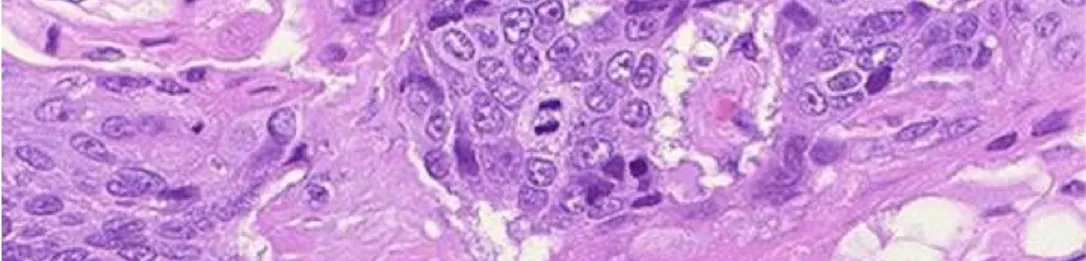
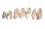 Staining — The tissues now need to be stained in order to see their microscopic details. Certain chemicals react differently with different tissues producing predictably varying colors. Hematolxylin, a blue basic dye, colors acid-containing materials blue. Because nuclei, for example, contain nucleic acids (DNA and RNA), these structures are colored blue. Eosin, a red acid dye, colors base-containing materials red. Collagen, a common component of oral tissues, has base units that stain red. Using hematoxylin and eosin (H & E) in staining has become the standard procedure for viewing tissue samples. Since hematoxylin is water-soluble and eosin is alcohol-soluble, it is necessary, after removing paraffin with xylol, to replace xylol to rehydrate prior to staining with hematoxylin and then again dehydrate prior to staining with eosin.
Adding Cover Slips — The stained sections are again placed in xylol to remove alcohol and allow for addition of “glue” (mounting medium) for affixing a very thin piece of glass over the tissues. The mounting medium must allow light to pass and, at the same time, firmly attach the cover slip. The finished slides are again placed in the incubator for sufficient time to allow the mounting medium to dry. Once sufficiently dry for handling, our pathologists can now examine the tissues from our submitting doctors.